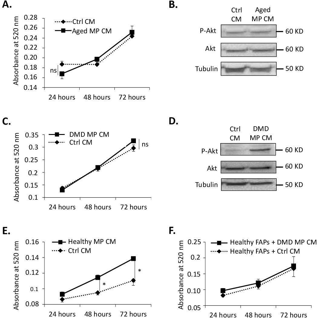
Fig. 2. The MP-dependent regulation of FAP proliferation is altered in aging and DMD progenitors. (A and B) Aged FAPs proliferated with control conditioned medium (Ctrl CM) or conditioned medium from aged MPs (Aged MP CM). (A) Proliferation was measured by MTT assays 24 hours, 48 hours and 72 hours after plating. (n=4 aged donors). (B) 15 minutes after addition of the conditioned medium, total proteins were extracted; expression levels of phosphorylated Akt, total Akt and tubulin were assessed by western blot. Representative immunoblot is shown (n=3 aged donors). (C and D) DMD FAPs proliferated with control conditioned medium (Ctrl CM) or conditioned medium from DMD MPs (DMD MPs CM). (C) Proliferation was measured by MTT assays 24 hours, 48 hours and 72 hours after plating. (n=6 DMD donors). (D) 15 minutes after addition of the conditioned medium, total proteins were extracted; expression levels of phosphorylated Akt, total Akt and tubulin were assessed by western-blot. Representative immunoblot is shown (n=3 DMD donors). (E) DMD FAPs proliferated with control conditioned medium (Ctrl CM) or conditioned medium from Healthy MPs (Healthy MP CM). Proliferation was measured by MTT assays 24 hours, 48 hours and 72 hours after plating. (n=3 DMD and n=1 healthy donor). (F) Healthy FAPs proliferated with control conditioned medium (Ctrl CM) or conditioned medium from DMD MPs (DMD MP CM). Proliferation was measured by MTT assays 24 hours, 48 hours and 72 hours after plating (n=3 DMD and n=3 healthy donors). ns= non-significant. * P < 0.05.